Scientific Program

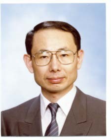
Koichi Shimizu
Waseda University, Japan
Title: Macroscopic 3D transillumination imaging of animal body by scattering suppression of NIR light
Biography:
Koichi Shimizu received M.S. (1976) and Ph.D. (1979) degrees, from University of Washington, Seattle, USA. He was Research Associate in University of Washington in 1974-79. He was Assistant Professor, Associate Professor, and Professor in Hokkaido University, Sapporo, Japan in 1979-2016. He is currently a Professor in Waseda University, Kitakyushu, Japan. He served as an associate editor of IEEE Trans. ITB in 1999– 2007. He is a Fellow of the Electromagnetics Academy, and an editorial board member of Scientific Reports (Nature).
Abstract
It has been widely known that we can see the surface blood vessel network of the thin part of our body. The near-infrared (NIR) light in the biological window range (700-1200 nm wavelength) is commonly used in biomedical applications such as a vein viewer and the vein authentication. The NIR light can penetrate much deeper in our body than the light of visible wavelength. However, the deep structure could not be visualized because of the strong scattering in body tissue. We developed some techniques to suppress the scattering effect and verified their feasibility in experiments. They are: the extraction of the near- axis scattered light (NASL) and the weakly scattered light (WSL) from the strongly scattered light. The deconvolution with a depth- dependent point spread function is another technique to suppress the scattering effect in a transillumination image. With these techniques we can visualize the macroscopic structure of an animal body in two dimensional (2D) images. Using the 2D transillumination images taken from different orientations, we can reconstruct the cross-sectional images and eventually the macroscopic three-dimensional (3D) image of the internal structure of an animal body. The effectiveness of the proposed technique was examined in the experiments with 3D model phantoms, and its applicability to an animal body was verified in the imaging of experimental animals.
- Optics and Biomechanics
- Modern Optics
- Interferometry
- Optical Metrology
- SDM and Beyond
- Optical Imaging Systems and Machine Vision
- Optical Computing
- Micro-Opto-Electro-Mechanical Systems (MOEMS)
- Microscopy and Adaptive Optics
- Lasers in Medicine and Biology
- Engineering Applications of Spectroscopy
- Applications and Trends in Optics
- Optoelectronic Devices, Photonics, Nanophotonics and Biophotonic
- Organic Optoelectronics and Integrated Photonics
- Optical Communications, Switching and Networks
- High-speed Opto-electronic Networking

