Scientific Program

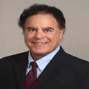
Robert W Thatcher
Applied Neuroscience Research Institute
USA
Title: New advances in electrical neuroimaging to evaluate EEG sources and brain networks
Biography:
Robert W. Thatcher, Ph.D., is currently the Director of Applied Neuroscience Research Institute and Applied Neuroscience, Inc. St. Petersburg, Florida. Dr. Thatcher is certified as an expert in both conventional electroencephalography and quantitative electroencephalography (QEEG), has read over 20,000 EEGs, and has written or supervised the writing of over 10,000 clinical EEG cases. He has extensive mathematical and programming experience as well as organizational leadership skills. He is the author of over 200 publications, including eight books. His most recent book is entitled the "Handbook of Quantitative Electroencephalography and EEG Biofeedback".
Abstract
The 3-dimensional evaluation of the sources of nonictal discharges and focal gross pathologies has recently been enhanced using advanced technology called swLORETA (weighted sLORETA. swLORETA uses Single-Value-Decomposition (SVD) to weight the lead field in order to increase lead field homogeneity and hence improved localization of deep sources. This allows for estimates of EEG sources in different layers of the cortex. Also, swLORETA uses a real MRI and not an average MRI with 12,270 voxels and a Boundary-Element-Method (BEM) of source localization (Wroel and Aliahadi, 2002). Non-ictal events and gross pathologies are localized inside of 3 dimensional volumes with the aid of slice and volume cutting tools to allow one to navigate through the brain and identify dysregulated brain network hubs (Brodmann areas) and connections. Computations include Functional Connectivity (Coherence, Lagged Coherence and Phase Difference) and Effective Connectivity (Phase Slope Index) of the magnitude and direction of information flow between network hubs as well as integration with Diffusion Tensor Imaging (DTI). A useful method is to also view the EEG potentials on a transparent scalp while simultaneously viewing the deeper sources of the EEG from inside the brain. Both raw scores and Z scores are used as well as the Laplacian transform of the scalp EEG. Examples of source localization in patients with traumatic brain injuries, strokes and epilepsy will be presented.
- Neuroscience & Therapeutics
- Developmental & Evolutionary Neuroscience
- Central nervous system
- Neuroanatomy & Physiology
- Behavioural neuroscience & Neurophysiology
- Clinical neuroscience & Neurosurgery
- Cellular Neurology & Neuroplasticity
- Molecular neuroscience & Neural engineering
- Neuroimaging & Neuroinformatics computational neuroscience
- Neurological disorder
- Neuropharmacology
- Pediatric Neuromuscular Disorders
- Neuro-oncology
- Neurogenetics
- Neurogastronomy
- Neuroheuristics & Neuroethology

