Scientific Program
Keynote Session:
Title: Fluidic-micro-nano-hetro-structures for nuclear power application
Biography:
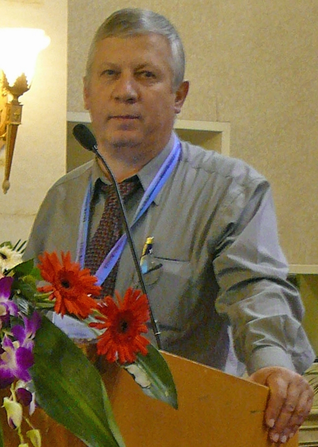
Popa Simil Liviu is a Nuclear Engineer, Physicist, Director of Los Alamos Academy of Sciences. He got B.Sc. In fast breeder reactors, M.Sc. In nuclear engineering, plasma-laser selective chemistry, and phd in nuclear, atomic, and molecular materials physics at National Institute for Atomic Physics, Bucharest.
Abstract:
Nuclear energy development is dependent on new nanoengineered materials incorporating heterogeneity by design, in order to allow higher performances and safety. Fission products occurrence inside nuclear reactors are responsible for low nuclear fuel burnup and fuel damage. Replacing the actual homogeneous Urania ceramic structure with a Ceramic –liquid metal micro structure that self-separates the fission products by trapping them into a drain liquid that may be NaK, PbBi (LBE) preventing them to damage the nuclear fuel and allowing easy fuel reprocessing. Another development relies on nano-cluster properties of enhanced separation of transmutation products resulted from Th or Pu cycle, or production of radioisotopes, that can be used inside nano-clustered-hetero-structure, made of nanobeads of actinides washed by a drain liquid with affinity for the transmutation products resembling a frit. Washed by the nanoflow. In direct nuclear energy to electricity conversion supercapacitor-like devices, the end of range of the nuclear particles create large lattice damage , that can be improved using liquid –solid hetero-structures as LiH on MWCNTs that have increased robustness to radiation damage. and sensitive lines.
Title: Recent investigations on microfluidics and nanofluidics using electrokinetics
Biography:
Ruey Jen Yang is a Chair Professor of Engineering Science at National Cheng Kung University (NCKU) in Taiwan. He received his phd Degree in Mechanical Engineering from University of California at Berkeley in 1982. From 1982 to 1993, he was affiliated with Rocketdyne in Los Angles working on the Space Shuttle Main Engine. He has been with the NCKU since 1993 after 11 years’ service in USA. His past academic services include Department Chair, Director General of Research and Services Headquarters and University Librarian at NCKU. His research interests include Microfluidics/Nanofluidics, Fluid and Thermal Sciences, Computational Physics, Energy and Nanotechnology. He has published more than 140 papers in archival Journals.
Abstract:
In this presentation, I will introduce our recent results: (1) utilizing fluidic technology with electrokientic effects to detect bio-samples and (2) preconcentrations by electrokinetic transport in nanospaces. Examples are using paper-based analytical devices (µpads) to detect Albumin, Creatinine, Glucose, Aspartate Aminotransferase (AST) and Alanine Aminotransferase (ALT) from human blood. The integrated system to perform these tests will be introduced in a portable platform.
Oral Session 1:
- Microfluidics Research and Advances | Microfluidic Chip | Lab-On-A-Chip Technology | Clinical Diagnostics

Chair
Prem Bikkina
Oklahoma State University, USA
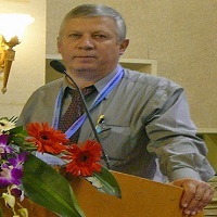
Co-Chair
Popa Simil Liviu
Los Alamos academy of Sciences, USA
Title: Microfluidics for particle generation: From a nanoparticle to a millimetre; from a milligram to a ton
Biography:
Richard Gray is a founder and director of Blacktrace, the group which includes two leading microfluidics companies - Dolomite and Syrris. He is responsible for US operations, and also Head of Sales for Dolomite Bio, a new group focusing on microfluidics products for biologists. An engineer by training, Richard graduated from Cambridge University and has worked in research and product development in aerospace, consumer products, healthcare and automated drug discovery.
Abstract:
Microfluidics offers a way to generate consistent, monodisperse particles in a wide range of materials. Almost any type of particle can be produced by these methods: from liposomes to multiple emulsions; from hydrogels to polymers. However, these methods have generally been limited to low throughput, and been more suited to lab-scale development work. Dolomite Microfluidics is addressing this limitation by focusing on higher throughput techniques and highly parallel approaches. We have developed new microfluidic methods and real time quality control tools, and we can now cover the range from a lab-scale development tool to ton-scale production-scale systems. This talk will explain our approach and give examples from a range of application areas.
Title: A centrifugal-based nucleic acid test with the immiscible filtration technique
Biography:
Chih-Hsin Shih has completed his phd from the Ohio State University. He is the associate professor in the department of chemical engineering, Feng Chia University, Taiwan.
Abstract:
A nucleic acid test (NAT) is a technique used to detect a particular nucleic acid, virus, or bacteria. In recent years, it has been applied to many applications such as clinical diagnoses, species identification, and food safety. However, in the conventional testing operation, the testing procedure for NAT is usually labor-intensive and time-consuming. It also required well-trained technician and the transfer of testing sample could result in nucleic acid contamination. Therefore, a user-friendly and automated assay process to conduct NAT is of great importance.
The major assay process for the NAT includes extraction, amplification, and detection. In the past few years, many microfluidic functions were developed for sample preparation and process automation. Kido et al. developed a cell-lysing approach by grinding the cell with beads. To control the movement of magnetic beads, Strohmeier et al. developed a Gas-phase transition magnetophoresis to transfer the magnetic beads through the liquid/air interface. To purify the nucleic acid, Berry et al. proposed an immiscible filtration assisted by surface tension technique.
In this work, an integrated and automated nucleic acid testing platform was developed using the centrifugal microfluidics. The microfluidic disk is able to lyse the cells, purified the nucleic acid through immiscible phases, and eluted the nucleic acid from the complex into the elution buffer. The eluted buffer can be used for amplification and quantification. A magnetic module was developed to transfer magnetic beads in the above-mentioned processes. This work provides an automated and user-friendly solution for the nucleic acid testing
Title: The development of a pico litre sample delivery system for XFEL time resolved studies
Biography:
Peter Docker holds the position of Senior Engineer at the Diamnd light source the UK’s synchrotron. He has 19 years experience in micro, nano and pico device and sample handling systems. His current positon involves developing such systems for sample delivery for Xray interrogation.
Abstract:
This paper describes new and exciting applications for a technology developed by Poly Pico ltd. We describe how this pico litre sample handling system is being developed for time resolved pico litre protein crystallography investigations. The studies are planned for LCLS II at Stanford University and the European XFEL in Hamburg.
The standard Poly Poco system can deliver discreet droplets of fluid or in our case, fluid containing nano dimensional protein crystals at a rate up to 10KHz. The system currently being developed will deliver at a rate up to 50KHz and can be externally triggered so sample can be delivered when pulses of X rays are present and timed with the detector rate. Current sample delivery systems deliver a jet of protein crystals continuously and data collection is only a few percent from the total crystals supplied. Our new approach is even more important when using X-rays at the European XFEL as their X-rays are delivered with an unusual time structure. The beam has a repeating signature repeating every 10HZ. It is off for 98% of the time and then delivers up to 2700 pulses before going dark again. These discrete pulses are ideally positioned to study chemistry reactions in real time. For clarity, the pico system could use 16 of these tightly packed pulses, the only system in the world capable of doing so. The Poly Pico system almost eliminates the sample wastage that is synonymous with continuous jetting. Other developments will also be described including temperature control for delivering viscous material and alignment using electro-steering.
Title: Generation of in vitro 3D hepatic tumor model for discovery of novel therapeutics
Biography:
Ting-Yuan Tu joined the Department of Biomedical Engineering at National Cheng Kung University (NCKU) in 2016 as an Assistant Professor. His phd work was completed in Singapore-MIT Alliance for Research and Technology Centre in Singapore, and received the Phd degree in Mechanobiology from National University of Singapore in 2015. Prior to joining NCKU, he was an Application Scientist at Clearbridge Biomedics (Now Biolidics). His current research interest lies in the development of better biomimetic tumor microenvironment for cancer drug discovery, such as in vitro tumor models and 3D tumor invasive co-culture microfluidic platforms.
Abstract:
In vitro 3D tumor model has been sought to mimic critical in vivo pathological features that could be used for discovery of therapeutic treatment. Fabricating microwells based on existing lithography/cleanroom-based approaches are often costly and time-consuming that requires laborious process. Based on our previous work, this study presented a rapid and economical microwell fabrication methodology aimed to be conveniently incorporated with conventional workflow from biological and medical laboratories for generating in vitro 3D tumor spheroids. Ablation of CO2 laser system was capable of easily incorporating z-axis adjustment to generate microwells with wide-rage size flexibilities, the side view of microwells revealed the depth and width fabricated under different laser power and focusing plane. Evaluation of tumor spheroid generated from different size of microwells was performed by CMFDA live cell staining, and hoechst nuclear staining indicated good cell viability and illustrated the shape of the tumor spheroids harvested from different size of microwells. To evaluate the preliminary therapeutic response on in vitro hepatocarcinoma tumor spheroid, photo-thermal treatment through bound ConA-Silicon Carbon Hollow Spheres (SCHS) was investigated, live/dead cell staining on tumor spheroid showed the photo-thermal effect could induce cancer cell death via exposure to NIR laser, resulting in higher red fluorescence under photo-thermal therapy. Preliminary therapeutic response from the hepatic 3D tumor spheroids with binding of ConA-SCHSs suggesting the current methodology combining this in vitro tumor model could serve as an effective tool for discovery of therapeutic motilities for cancer treatment.
Title: Manipulation of biomimetic soft interfaces by optical and microfluidic methods
Biography:
Guido Bolognesi obtained an international joint PhD in “Theoretical and Applied Mechanics” in 2012 at the University of Rome “La Sapienza” and University Claude Bernard Lyon 1 (UCBL). In 2011, he joined the Membrane Biophysics Group in the Department of Chemistry at Imperial College London as a post-doctoral research associate and in 2016 he became lecturer in Bio-engineering in the Department of Chemical Engineering at Loughborough University. His research interests lie in the experimental investigation of the physico-chemical behaviour of soft matter (such as colloids, lipid membranes) and fluid flows at the micrometer length scale with an interdisciplinary approach based on expertise in mechanics, micro-/nano-fluidics, microfabrication techniques, optics, interface and colloid science.
Abstract:
The design and manufacturing of functional soft structures, based on multiple compartments delimited and interconnected by lipid-stabilised or surfactant-stabilised interfaces, are attracting an increasing level of attention. These soft constructs have shown a great potential as minimal cells in synthetic biology, simplified model systems for biophysical and biochemical studies and smart containers for drug delivery and microreactor technologies.
In recent years, microfluidic and optical methods for the manipulation of such soft interfaces have provided a substantial contribution to the design and development of novel artificial systems exhibiting interesting cell-like behaviours and functions. In this talk, I will present a number of optical and microfluidic techniques we developed for the construction and characterisation of artificial soft structures, including droplet interface bilayer architectures and soft nanotube networks. More specifically, I will discuss how accurate flow and particle handling operations within a microfluidic environment can be used to build artificial model systems from soft interfaces, such as lipid bilayer membranes and surfactant-stabilised oil-water interfaces. The resulting architectures can display biochemical and structural properties and functionalities, similar to those seen in living cell systems.
Title: Streamlined drug synergy evaluations via bioinspired nanodroplet processing platform for optimal cancer treatment
Biography:
Ching-Te Kuo received the phd degree in Institute of Applied Mechanics from National Taiwan University, Taipei, Taiwan, in 2013. In 2013-2018, his Postdoctoral Studies, from National Taiwan University to UT Southwestern Medical Center, focused on the development of in vitro mimicking of tumor microenvironments as well as high-throughput drug screenings by microfluidics. Currently, he is a Postdoctoral Researcher at National Taiwan University. His research interests include microfluidics, system control engineering, bioinspired material, tumor microenvironment and metastasis.
Abstract:
Synergistic combination of two or more drugs has been a major avenue targeting cancers. This regimen not only improves the therapeutic efficacy by triggering synthetic lethality in target cells but also minimizes the side effects by reducing drug doses. Therefore, the identification of optimal combination of various possible concentrations from a set of drugs presents a substantial challenge. Several approaches to optimize the selection regime have been demonstrated by various approaches; however, challenges still remain. For example, the time adopted for cell/drug preparation among the total independent screenings would last for days. In addition, the usage of standard multi-well plate assays would counter the feasibility for personalized medicine, which is inherently subject to a limited cell count from patient tumors. To address the technology gap described above, we herein present a bioinspired nanodroplet processing (BioNDP) platform for facilitating the high-throughput screening of optimal drug combinations. The platform was fabricated by a novel wax-imprinted laser direct writing, which is inspired from Stenocara gracilipes beetle’s bumpy back surfaces. Leveraging on advantages of utilizing customized liquid dispensing, cell counts and drugs adopted can be retained down to 50 cells and 200 nl for each test, respectively. We demonstrated an approximate 500-fold miniaturization of drug volumes does not impact both the in vitro and in vivo outcomes. In addition, such platform could present in vivo predictions more accurate than standard drug screening assays. Taken together, our results highlight the BioNDP platform could serve a cost-effective and high-throughput toolkit for improving pre-clinical drug screenings.
Title: Inertial micromixing in curved serpentine micromixers with different curvature angles
Biography:
Rana Altay is studying in Sabanci University, Turkey.
Abstract:
Micromixing is a crucial component of microfluidic systems which require mixing reagent molecules, fluids, or species for chemical reactions, which has applications in biomedical systems, chemical reactors, and polymerization . In this study, three curved serpentine micromixers consisting of ten segments with curvature angles of 180°, 230°, and 280° were fabricated to investigate the effects of curvature angle on inertial micromixing of two fluids. In this regard, water and diluted Rhodamine B solution were pumped into the micromixers over flow rates of 400-3000μL/min. To characterize and compare the mixing performance of the micromixers and to understand the underlying mechanisms, fluorescent intensity maps and mixing indeces were utilized. According to the results, up to the Reynolds number of 150, the mixing performance of the micromixers with curvature angles of 180° and 230° was similar to each other. While the micromixer having segments with 280° curvature angle showed higher mixing index values and thus outperformed the other two micromixers. This was due to severe distortion of flow streamlines by Dean vortices and occurrence of chaotic advection.
Title: Passive microfluidic production of multiple emulsions
Biography:
Adam S. Opalski is last year graduate student at Institute of Physical Chemistry of Polish Academy of Sciences in group of prof. Garstecki. His research interests are passive droplet microfluidics and multiple emulsions. He is funded by National Centre of Science of Poland.
Abstract:
Double emulsions (DE) are complex fluid systems, with a core droplet (e.g. aqueous) surrounded by a liquid shell (e.g. of oil) and suspended in an external phase (e.g. aqueous).The thinner the shell is, the higher stability is shown by the DE. They found use in various fields of life sciences, from production of polymerosomes to screening rare cells via flow-cytometry.
Microfluidic production of emulsions is thoroughly investigated, both single and complex emulsions. Double emulsions, however, are mainly produced by active methods, i.e. by shearing off a core droplet (creating feed, single emulsion) and shearing off the feed emulsion into double emulsion. Passive methods, such as step emulsification, offer higher level of monodispersity of produced emulsions as well as greater ease of use and parallelization than active methods.
Keynote Session:
Title: Automated venting technique used in microfluidic digital logic lab-on-a-chip device
Biography:
Manasi Raje is a microfluidics R&D Engineer working in Bay Area, California. She is working at Shield Diagnostics and developing diagnostic devices for affordable testing of infectious diseases. She holds training in Biomedical Engineering and Electronics Engineering. Manasi has experience in the design and fabrication of a variety of lab-on-a-chip devices. She developed a number of application specific and high-throughput-attempting micro fabrication methods during her time at JBEI that let to a patent.
Abstract:
Microfluidic digital logic is an emerging technology which can be useful to automate a variety of lab processes in order to bring lab tests and diagnostics to low resource settings where it can be difficult to establish expensive and advanced labs. Microfluidic digital logic derives its concept from the digital logic in electronics and aids to automate complex liquid handling processes. The basic logic circuits are designed using valves and pneumatic resistors in a way that can be used to pump fluid around in the chip in order to transport the fluid or to mix it or store it. Automation of such basic aspects of a microfluidic device can help to eliminate the wieldy machinery that is required to drive a microfluidic chip. For example the computer program, mechanical/electrical pumps and syringe pumps can be replaced by on-chip pneumatic logic circuits. The heart of this work is a serial dilution circuit which has been previously reported in detail. The logic blocks designed around the dilution ladder do the job of selecting the program, pumping the diluent and making sure all the logic signals are routed correctly without any leakage or impeding attenuation. The circuit that receives and routes all other signals incorporates a feature to check the quality of dilution, the feature is called the venting network.
Title: The state-of-the-art of surface modification techniques for geo-material microfluidics
Biography:
Prem Bikkina is an Assistant Professor in the School of Chemical Engineering, OSU, Stillwater. He has B.S. and M.S. degrees in chemical engineering and phd degree in petroleum engineering. He worked as a postdoctoral fellow at Lawrence Berkeley National Laboratory. He also worked in various chemical and petroleum industries. His research work on enhanced hydrocarbon recovery, geological sequestration, and multiphase separation resulted into high impact journal publications and patents. He has been a peer reviewer for more than 16 international journals, ACS PRF and DOE proposals. Dr. Bikkina received ‘2016 Outstanding Reviewer Award’ from the Journal of Environmental Chemical Engineering. He received ‘2016 SPE Mid-Continent Regional Service Award’ and ‘2017 SPE Distinguished Petroleum Engineering Faculty Award’. He is a professional member of SPE, aiche, ACS, and ASME.
Abstract:
In recent years, microfluidics has been gaining acceptance for the fundamental and applied petroleum engineering research. The major advantages of using microfluidics include flexibility in porous media and other related chip designs in a highly controlled and reproducible manner, easy and accurate control of fluid flow, and most importantly the ability to visually study the involved fluid distribution and displacement mechanisms both at pore and chip scales. The recent advancements in manufacturing techniques made it possible to prepare microfluidic chips that could replicate various pore-scale features of real porous media, and other patterns that are of relevance to petroleum engineering applications. However, a major limitation for the application of microfluidics for subsurface multiphase fluid flow and reactive transport is the dissimilarity in the physicochemical aspects of the real porous media and microfluidic solid surfaces. This presentation discusses the state-of-the-art of the surface modification techniques currently being used in geo-material microfluidics.
Oral Session 1:
- Bio-MEMS/NEMS and Chips | Biosensors, types and Bio sensing | Fluidic Micro Actuators | Microfluidics in Nano-medicine
Title: Plasma effects on microbubble formation in gas-liquid interface across a microfluidic plasma reactor
Biography:
Oladayo Ogunyinka has completed his masters degree in mechanical engineering at the age of 22 years from the University of Sheffield, UK. He is currently a research student in microfluidics and plasma field at Loughborough University, UK.
Abstract:
Atmospheric-pressure plasmas has increasingly been studied for their potential in many applications, such as water treatment, generation of reactive species, biological applications, and material synthesis. This study involves the use of atmospheric-plasma in microscale to transfer plasma reactive species to organic liquid via microbubbles gas-liquid interface. Hence, plasma interaction with microbubbles is a topic to investigate.
Title: Biosynthesis of zinc oxide nanoparticles using Albizia lebbeck stem bark and evaluation of its antimicrobial, antioxidant and antiproliferative activities on human breast cancer cell lines
Biography:
Huzaifa Umar has completed his Masters at the age of 26 years and PhD Studies at the age of 32 from Cyprus International University, Lefkosa, Turkey. He is the assistant director, biotechnology research center, Cyprus International University. He has published more than 15 papers in reputed journals.
Abstract:
Biocompatibility and stability of zinc oxide nanoparticles (ZnO NPs) synthesized using plants is a promissing research area of study in nanotechnology, due to its wide applications in biomedical, industrial, cell imaging and biosensor fields. The present study reports the novel green synthesis of stable ZnO NPs using various concentrations of zinc nitrate 0.01, 0.05, 0.1 M, and Albizia lebbeck stem bark extracts as an efficient chelating agent. Antimicrobial, antioxidant, cytotoxic and antiproliferative activities of the synthesized nanoparticles on human breast cancer cell lines, were evaluated using different assays. The UV-vis spectroscopy result revealed an absorption peak in the range of 370 nm. The involvements of A. lebbeck bioactive compounds in the stabilization of the ZnO NPs was confirmed by X-ray diffraction (XRD) and Fourier transform infrared (FTIR) analysis. Zeta sizer studies showed an average size of 66.25 nm with poly disparity index of 0.262. Scanning electron microscope (SEM), Energy-dispersive X-ray spectroscopy (EDX) analysis results, revealed irregular spherical morphology and presence of primarily Zn, C, O, Na, P and K respectively.

