Scientific Program
Keynote Session:
Title: Outcome of neglected elbow dislocation treated with open reduction at rural tertiary care hospital in Nepal
Biography:
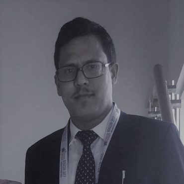
Mangal Rawal has completed his bachelor in medicine from B.P. Koirala Institute of Health Science, Dharan, Nepal. He has completed master in orthopedic surgery from National Academy of Medical Sciences, Kathmandu. He also has completed his fellowship in trauma and illizarov from National Institute of Trauma and Rehabilitation (NITOR), Dhaka, Bangladesh. Currently he is working as a head of department of orthopedics and trauma at Karnali Academy of Health Sciences which is one of the pioneer tertiary care institution in rural Nepal. He is taking a responsibility as a health policy advisor of provincial government of Karnali province.
Abstract:
According to many kinds of literature published, the old unreduced dislocation of the elbow is a rare condition. But it seems more frequent in developing countries due to lack of awareness among the patient population who believe in traditional faith healers and lack of proper health facilities. Our institute is located in the most rural parts of Nepal so that the incidence of neglected dislocation of elbow might be more common than other parts. Neglected elbow dislocation has a great impact on the activity of daily living. Open reduction of neglected elbow dislocation, most of the time, leads to less than expected outcome. There are several methods by which neglected dislocation can be treated. We have compared the functional outcome of 11 cases of neglected dislocation treated with open reduction and trans olecranon K wire fixation over a period of 3 years.
Material and methods: 11 consecutive cases of neglected elbow dislocation presented to our institute were evaluated and treated with open reduction and K wire fixation. The functional outcome was measured with MEPI scores. Preoperative and postoperative extension, flexion and range of motion were evaluated. Associated surgical complications were also studied.
Results: 7 patients had a history of fall from height and 4 patients presented with slipped fall on the ground. Average time of presentation after trauma was 6.18 month. The average age of the patient was 28.2 years. MEPI has increased from 15.45 preoperatively to 87.27 postoperatively. No major complication associated with the treatment except one case of the unstable elbow.
Conclusion: Open reduction and K wire fixation provide a good functional outcome in a neglected elbow dislocation of more than 3 weeks duration.
Title: Management of postoperative pain in total knee arthroplasty patients: A clinical overview
Biography:
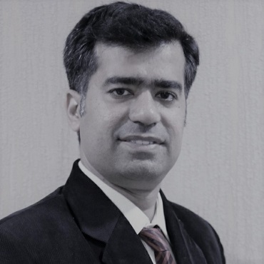
Kunal Makhija has completed his Masters from Asia largest hospital B J medical hospital and acquired sub specialty in Subvastus Knee replacement from Telford, UK and Bombay Hospital, India. He has International as well as National publications and presentations. He is the Director of Joint clinic, India.
Abstract:
Total knee arthroplasty has become a household surgery in recent times with innovations happening every day. Failure to achieve pain relief in immediate post operative period causes sleep disturbances, rehabilitation delay, mental stress, delayed discharge and long term post surgical discontent1.In Era of day care surgeries and robotics, post operative comfort of patient needs to be utmost important. A combination of pain relieving measures needs to be taken rather than single approach. The goal of this article is to understand the fundamental concepts of surgical pain, understand the molecular mechanism of agents and to use this knowledge to provide best possible pain relief measures. The neurogenic and nociceptive pain relieving agents were used in pre defined combination and dosage in 200 patients and pain, stay and rehabilitation was observed. A combined preventive analgesia approach starting two days before surgery and lasting upto 24 hours after surgery is effective method to control post operative pain.
Title: Is there a need for a fellowship-advisor for orthopaedic surgeons? Analysis of 543 surveys from my-fellowship.com, a Swiss online platform
Biography:
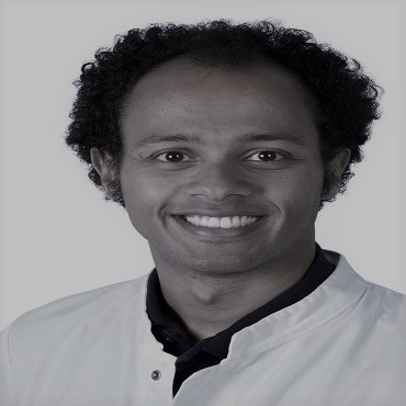
Mohy Taha is a board-certified and fellowship trained orthopaedic surgeon in Basel-Switzerland, with extensive training in shoulder and elbow surgery. He is an author, a speaker and the founder of My-Fellowship.com. He finished his medical school in Egypt, accomplished his training in Switzerland and completed 5 fellowships and 9 Observer ships in Australia, Brazil, Canada, France, Germany and USA. In 2013, he was looking for fellowships to further his training. He visited 13 clinics in Australia to find a suitable fellowship that matches his training needs. During his fellowship in 2016 he met some peers who were not satisfied with their fellowships. The idea of a platfrom that can bridge the gap between fellowship offers and fellows' expectations was born.
Abstract:
Introduction: Fellowships are common in many countries, whereas other countries do not offer special training after residency. Therefore many doctors seek for a fellowship after completing their residency. Fellowships became an essential part of professional medical training. Finding a suitable fellowship is essential. Physicians and institutions may have different expectations regarding fellowships, which can lead to frustration and wasted resources for both. One obstacle for doctors is finding reliable feedback from previous fellows regarding a specific fellowship and being able to contact that person for further advice. In addition, for both doctors and institutions alike, financing a fellowship can also prove a challenge.
Methods: A website was created as well as a 1 minute survey. The project was initiated during an International Orthopaedic and Trauma meeting in Switzerland. Moreover, the website was posted frequently on Linkedin, Facebook, Instagram, Twitter and a newsletter explaining the project was sent to 1749 doctors worldwide per e-mail.
Results: 3.5 months after the project initiation there were 6396 visits (5287 new users) to the website from 124 countries, mainly USA (31.3%), Switzerland (20.4%), Egypt (5.5%) and India (4.2%). 543 surveys from participants from 72 countries were completed: Switzerland (25.4%), Egypt (7.4%), India (6.6%) as well as United Kingdom (5.5%), United States (4.4%), Colombia (4.1%) and Brazil (3.9%).
The main specialties were Orthopaedic & Traumatology (61.8%), General Surgery (8.1%) and Internal Medicine (4.1%). Most of the participants were potential fellows (38.5%), previous fellows (31.7%) or individuals and/or institutions who would like to offer a fellowship (17.9%). The participants were mainly interested in a fellowship database (72.6%), connecting to other fellows (65.4%), and giving/receiving feedback about a fellowship (51.9%) or receiving financial support (37.9%).
Conclusion: The results of the first surveys suggest that there is an interest in an online fellowship advisor including a database for fellowships worldwide, a platform for fellows to connect to each other with the ability to give and receive feedback about a fellowship.
Accordingly an IT company was assigned to build the platform with the needs of doctors and institutes in mind. Financing a fellowship remains a challenge for many participants.
Title: Osseointegrated, percutaneously derived implants for rehabilitation after transfemoral amputation - via the Endo-Exo Implant System
Biography:
Marcus Oergel currently working as professor at the Trauma Department, Hannover Medical School, Germany.
Abstract:
Introduction: Osseointegrated, percutaneously guided implants - so called endo-exo prosthetics (EEP) - have been used to a limited extent since 1999 for rehabilitation after major amputation. Meanwhile, implant survival times of more than 15 years have been achieved. The obligatory colonization of the skin penetration site of the implant does not necessarily lead to an intramedullary, periprosthetic infection. This circumstance can be explained by the interconnectivity ingrowth of the bone into the three-dimensionally structured implant surface. This sufficiently prevents the formation of an infection-promoting connective tissue layer between bone and metal.
Material/Method: A total of 110 EE femoral prostheses were implanted (6 x bilaterally amputated) in 104 patients between August 1999 and October 2016. The implant is produced by casting from a CoCrMo alloy coated with titanium nitride. The first step is the implantation (Figure 1) of the intramedullary module with subsequent wound closure. After safe osseointegration of the endomodule after 6 weeks, the skin was penetrated (Figure 2) with docking of the components receiving the exoprosthetics in a second surgical step.
Results: The retrospective analysis shows that a total of 324 operations were required for the 110 implantations performed. Of these, 220 operations were performed using the two-stage implant procedure. The remaining 104 surgical interventions were soft tissue problems at the skin interface, 7 fracture restorations, 8 explantations with 3 reimplantations and minor corrections to the prosthetic components. The initial infection problem with severe soft tissue irritations at the skin interface could be effectively countered by changing the design of the components. From January 2010 - October 2016, only 12 surgical procedures were necessary. It has also been shown that even prolonged soft tissue infections involving the distal end of the femur do not necessarily lead to an ascending periprosthetic infection. Patient satisfaction was predominantly high. Regarding to the wearing and operating comfort of the prosthetics, the recovery of a tactile sensation, the so-called "osseoperception", and the considerable increase in mobility are these very positive aspects. When using the bone-guided prosthesis, special attention must be paid to the orthopaedic technical treatment. In particular, the axial alignment of the prosthesis abutment requires specific handling in order to achieve an optimal walking pattern and to prevent possible consequential damage to the hip joint and spine.
Conclusions: Bone guided percutaneous prosthetics for rehabilitation after transfemoral amputation can be considered sufficiently safe according to the available data. Therefore it represents a valuable treatment option for patients after transfemoral amputation who would otherwise not be able to rehabilitate satisfactorily.
Rohit Kulkarni
Consultant Orthopaedic Surgeon in Pune and is the Head of Department of Orthopaedics at Om Hospital, IndiaTitle: Medial opening wedge high tibial osteotomy with the navigated iBalance HTO system and early weight-bearing: Evaluation of precision and maintenance of correction
Biography:
Rohit Kulkarni has completed both his diploma and degree in orthopaedics from renowned institutes in India. He has completed board certified fellowships from Australia and which are accredited by the Australian Orthopaedic Association. He has international journal publications and has paper and poster presentations across various orthropaedic conferences in India. He is currently practicing as a Consultant Orthopaedic Surgeon in Pune and is the Head of Department of Orthopaedics at Om Hospital, Pune. He serves as a panel consultant in the Department of Orthopaedics at MJM Hospital, AIMS Hospital and Jupiter Hospital in Pune, India.
Abstract:
Purpose: Safety and stability of the Arthrex iBalance HTO system has been demonstrated using standard surgical techniques and conventional postoperative rehabilitation protocols. The purpose of the current study was to investigate the accuracy and stability of the osteotomy following iBalance implantation using a minimally-invasive, navigated surgical technique, in combination with an accelerated rehabilitation protocol.
Methods: A prospective, observational study of 20 consecutive patients undergoing medial opening HTO with the iBalance implant and a minimally-invasive, computer-navigated surgical technique was conducted. Intraoperative stressed hip-knee-ankle (HKA) angles measured with navigation were compared to 2 weeks postoperative HKA angles measured on long leg radiographs to determine the accuracy of the surgical technique. The maintenance of correction was assessed to 1 year postoperative. Time to union and full weight bearing and pre- and postoperative patient-reported outcome measures (PROM) were also evaluated.
Results: All knees were corrected to valgus, with the target correction of 2o to 3o valgus achieved for 63.2% of patients. No significant differences were observed between mean intraoperative stressed HKA and mean postoperative HKA angles. The lateral cortical breach occurred in one patient postoperatively; however, no additional complications arose throughout the study period. PROM demonstrated a significant reduction in pain scores and increased mobility between 6 weeks to 3 months postoperative. The mean deviation of correction was 1.4° ± 1.7° at 1-year post-surgery.
Conclusion: Intraoperative use of computer navigation was able to accurately reproduce pre-planned correction angles, with the maintenance of tibial correction over 1 year using the iBalance in combination with an accelerated rehabilitation program.
Title: Role of epidural steroid plus local anaesthetic in lumbar canal stenosis in older adults
Biography:
Swaraj Sathe is an orthopedic surgeon in Kothrud, Pune, India and has an experience of 9 years in this field. He practices at Sathe Multispeciality Hospital in Kothrud, Pune, and Ruby Hall Clinic in Hinjewadi, Pune. He completed MBBS from Krishna Institute of Medical Sciences, Karad in 2009, MS - Orthopaedics from Goa Medical College, Panaji in 2013 and DNB - Orthopedics/Orthopedic Surgery from Goa Medical College, Panaji in 2014. He is a member of Indian Medical Association (IMA), Indian Orthopaedic Association and Pune Orthopaedic Society.
Abstract:
Statement of problem: Lumbar canal stenosis (LSS) is one of the most commonly diagnosed and treated pathologic conditions affecting the spine. Prevalence of acquired, degenerative LSS ranges from 16.8 – 29.1%. 1 LSS causes significant disability in elderly. Epidural steroid with local anaesthetic is less invasive, safer, and more cost effective treatment than surgery.2
Methodology: 10 patients with evidence of LSS on MRI and significant neurological claudication and paraesthesias on history and clinical examination were selected. There was no motor deficit or bowel/bladder involvement. Patients were aged between 60 and 79 years; 3 patients were female and 7 were male. The patients were given triamcinolone with bupivacaine and lignocaine in epidural space at level of L2-3, L3-4. Patients with motor deficit, bowel/bladder involvement, and evidence of spinal instability on x-ray, prior lumbar surgery and prior epidural injection in past 6 months were excluded. Pain was classified according to Visual Analogue Scale. Patients were evaluated at 2 weeks, 4 weeks and 12 weeks.
Findings: 8 out of 10 patients reported significant improvement in pain, 1 reported temporary decrease in pain and 1 reported no improvement.
Conclusion and significance: Epidural steroid injection is an effective treatment to improve pain and function among older adults with LSS. It has the advantage of being a day care procedure, with less comorbidity and expenditure compared to surgery.
Title: Results of Antibiotic impregnated cement coated IM Nail in management of infected Non union and Compound fractures of long bones
Biography:
Pankaj Kumar Singh Ms (Orthopaedics) currently working as Senior Resident in Dept. Of Orthopaedics and Traumatology, IGIMS, India.
Abstract:
Aims and Objectives: The aim of study is to analyze the results of antibiotic impregnated cement coated IM Nail in management of infected nonunion and compound fractures of long bones (upto Grade 3A); to assess the effectiveness in eradication of infection, the rate of bony union and the functional return of the limb in the post-operative period.
Design: Prospective study.
Setting: Private Hospital Setup.
Intervention: Surgery.
Patients: A total of 25 patients were included in the study with infected nonunion and compound fractures of long bones (upto Grade 3A) for treatment with this method from August 2013 to May 2017.
Main outcome measurement: A minimum of 2.5 years follow up was done to assess the return of the functional outcome of the limb, rate of bony union (According to ASASMI criteria) and effectiveness in eradication of infection.
Results: There were excellent results in bony union at the fracture site (except in 3 cases) and excellent results in the functional outcome of the limbs. Control of infection was also achieved in most of the patients (except in 3 cases). Overall, the outcome following this treatment for infected nonunion and compound fracture of long bone was good to excellent.
Conclusion: In our study we found that bony union, infection control and functional results are good to excellent. Radical debridement with removal of all sequestrated bone fragment is mandatory before implantation of antibiotic coated IM nail in infected nonunion of long bones.
Title: Osseointegrated, percutaneously derived implants for rehabilitation after transfemoral amputation - via the Endo-Exo Implant System
Biography:
Marcus Oergel currently working as professor at the Trauma Department, Hannover Medical School.
Abstract:
Introduction: Osseointegrated, percutaneously guided implants - so called endo-exo prosthetics (EEP) - have been used to a limited extent since 1999 for rehabilitation after major amputation. Meanwhile, implant survival times of more than 15 years have been achieved. The obligatory colonization of the skin penetration site of the implant does not necessarily lead to an intramedullary, periprosthetic infection. This circumstance can be explained by the interconnectivity ingrowth of the bone into the three-dimensionally structured implant surface. This sufficiently prevents the formation of an infection-promoting connective tissue layer between bone and metal.
Material/Method: A total of 110 EE femoral prostheses were implanted (6 x bilaterally amputated) in 104 patients between August 1999 and October 2016. The implant is produced by casting from a CoCrMo alloy coated with titanium nitride. The first step is the implantation (Figure 1) of the intramedullary module with subsequent wound closure. After safe osseointegration of the endomodule after 6 weeks, the skin was penetrated (Figure 2) with docking of the components receiving the exoprosthetics in a second surgical step.
Results: The retrospective analysis shows that a total of 324 operations were required for the 110 implantations performed. Of these, 220 operations were performed using the two-stage implant procedure. The remaining 104 surgical interventions were soft tissue problems at the skin interface, 7 fracture restorations, 8 explantations with 3 reimplantations and minor corrections to the prosthetic components. The initial infection problem with severe soft tissue irritations at the skin interface could be effectively countered by changing the design of the components. From January 2010 - October 2016, only 12 surgical procedures were necessary. It has also been shown that even prolonged soft tissue infections involving the distal end of the femur do not necessarily lead to an ascending periprosthetic infection. Patient satisfaction was predominantly high. Regarding to the wearing and operating comfort of the prosthetics, the recovery of a tactile sensation, the so-called "osseoperception", and the considerable increase in mobility are these very positive aspects. When using the bone-guided prosthesis, special attention must be paid to the orthopaedic technical treatment. In particular, the axial alignment of the prosthesis abutment requires specific handling in order to achieve an optimal walking pattern and to prevent possible consequential damage to the hip joint and spine.
Conclusions: Bone guided percutaneous prosthetics for rehabilitation after transfemoral amputation can be considered sufficiently safe according to the available data. Therefore it represents a valuable treatment option for patients after transfemoral amputation who would otherwise not be able to rehabilitate satisfactorily.
Title: Cbfβ/Runx1 complex is important for articular cartilage integrity
Biography:
Currently she is Young Scientist Researcher at Kyungpook National University, Korea.
Abstract:
Osteoarthritis (OA), a leading age-related disease in society, still lacks a clear molecular mechanism. Here, we explored in vivo role of core binding factor β (Cbfβ) in OA by generating articular cartilage-specific Cbfβ-deleted mice (Cbfβâ–³ac/â–³ac) using Gdf5 promoter-driven Cre mice. OA was induced through destabilization of the medial meniscus (DMM) surgery in 12-week-old male mice. At 8 weeks after surgery, OA phenotypes were more accelerated in Cbfβâ–³ ac/â–³ ac mice than wild type (WT) mice with increased expression of Mmp13 and decreased expression of Type II collagen. Interestingly, the expression of Cbfβ was reduced during aging as determined by immunohistochemisty. Furthermore at 5 months of age Cbfβâ–³ac/â–³ac mice, but not in WT, exhibited OA naturally without developmental defects in joint and skeletal tissue formation. To explore the molecular mechanism of the protective role of Cbfβ in OA, we measured the expression of chondrocyte markers, Runx transcription factors, and Cbfβ in articular cartilage. Expression of chondrocyte markers such as type II collagen, Aggrecan, and Cbfβ was attenuated in chondrocytes derived from Cbfβâ–³ac/â–³ac OA mice compared to WT mice. Among Runx family, Runx1, but not Runx2 and Runx3, was highly expressed in particular chondrocytes. Expression of Runx1 was gradually decreased during OA progression in WT mice. Importantly, Runx1 expression was further diminished in Cbfβâ–³ac/â–³ac OA mice. Cbfβ formed a complex with Runx1 and protected Runx1 from proteosomal degradation in primary articular chondrocytes as well as in ATDC5 cells. Consistently, forced expression of Cbfβ in Cbfβ-deficient primary articular chondrocytes restored the chondrocyte markers and Runx1 expression. Collectively, these results demonstrate that Cbfβ is required for Runx1 stability as a partner protein in articular cartilage and that the formation of the Cbfβ-Runx1 complex plays an essential role for maintenance of articular cartilage integrity.

